
The ultrasound technique demonstrates a steep learning curve and requires detailed knowledge of the foot anatomy.
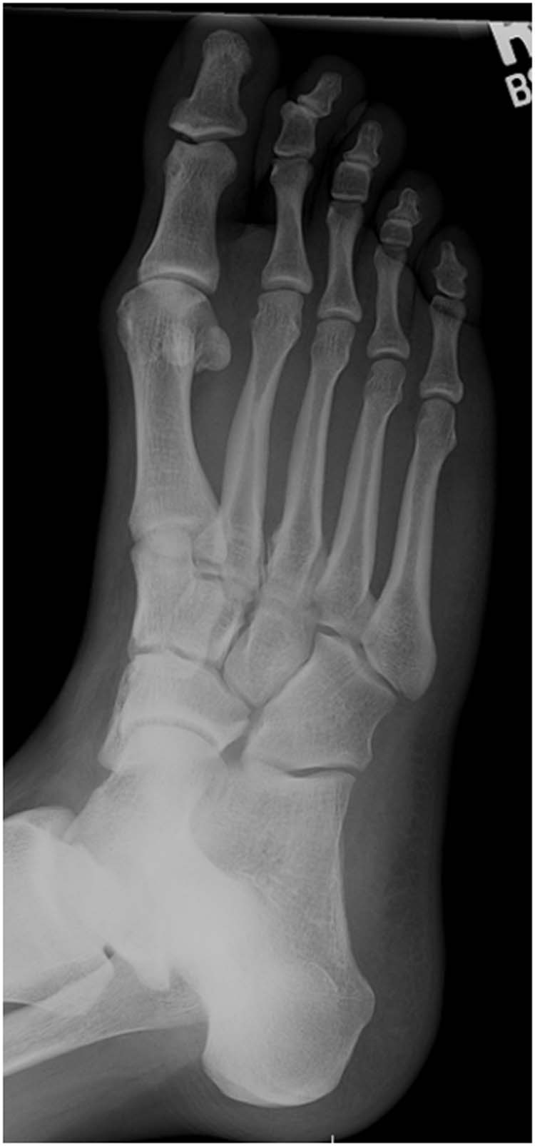
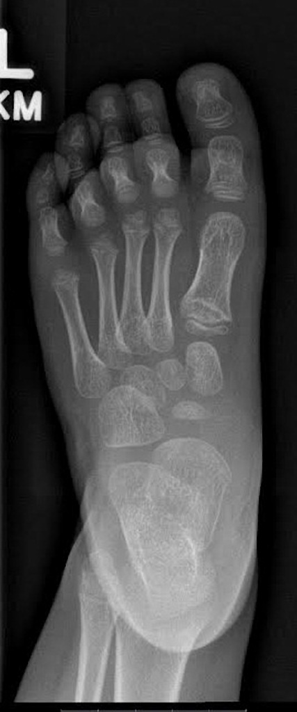
#Normal foot xray portable
High-frequency sonography is inexpensive, portable and is unique in allowing true dynamic assessment of the ligamentous, muscular and tendinous structures. J Foot Ankle Surg 1998 37(6):524–530.Traumatic, degenerative and rheumatological injuries of the foot are common and can be managed by an ever increasing number of treatments and surgical interventions. Joplin’s neuroma or compression neuropathy of the plantar proper digital nerve to the hallux: clinicopathologic study of three cases. Morton neuroma: MR imaging in prone, supine, and upright weight-bearing body positions. Weishaupt D, Treiber K, Kundert HP, et al. Medial calcaneal nerve: an anatomical study. The medial and inferior calcaneal nerves: an anatomic study. Variations in the origin of the medial and inferior calcaneal nerves. Branches of the tibial nerve: anatomic variations. Tarsal tunnel syndrome: a review of the literature. Tibial nerve branching in the tarsal tunnel. Havel PE, Ebraheim NA, Clark SE, Jackson WT, DiDio L. Sonographic imaging of the sciatic nerve division in the popliteal fossa. Schwemmer U, Markus CK, Greim CA, Brederlau J, Kredel M, Roewer N. Primal’s 3D atlas of human anatomy of the leg, ankle and foot. Risk of sural nerve injury with intramedullary screw fixation of fifth metatarsal fractures: a cadaver study. Donley BG, McCollum MJ, Murphy GA, Richardson EG. Ultrasound of the sural nerve: normal anatomy on cadaveric dissection and case series. Belsack D, Jager T, Scafoglieri A, et al. The bilateral anatomical variation of the sural nerve and a review of relevant literature. Vuksanovic-Bozaric A, Radunovic M, Radojevic N, Abramovic M. The deep peroneal nerve in the foot and ankle: an anatomic study. Anatomic variations of superficial peroneal nerve: clinical implications of a cadaver study. Prakash, Bhardwaj AK, Singh DK, Rajini T, Jayanthi V, Singh G. Sonography of the normal ulnar nerve at Guyon’s canal and of the common peroneal nerve dorsal to the fibular head. Ultrasound of the musculoskeletal system. High-resolution magnetic resonance imaging of the lower extremity nerves. Imaging of foot and ankle nerve entrapment syndromes: from well-demonstrated to unfamiliar sites. Delfaut EM, Demondion X, Bieganski A, Thiron MC, Mestdagh H, Cotten A. MR imaging of neuropathies of the leg, ankle, and foot. Allen JM, Greer BJ, Sorge DG, Campbell SE. Imaging of nerve entrapment in the foot and ankle. Peripheral nerve entrapments of the lower leg, ankle, and foot. Philadelphia, Pa: Wolters Kluwer–Lippincott Williams & Wilkins, 2011 381–427. Sarrafian’s anatomy of the foot and ankle. The authors’ discussion focuses on the superficial and deep peroneal, sural, saphenous, tibial, medial and lateral plantar, medial and inferior calcaneal, common digital, and medial proper plantar digital nerves. In addition, the authors illustrate proper probe positioning, which is essential for visualizing the nerves at US. The authors review the normal anatomy and common variants of the nerves of the foot and ankle, with use of dissected specimens and correlative US and MR imaging findings.

It may be difficult to distinguish the nerves from adjacent vasculature at MR imaging, and US can help in differentiation. Ultrasonography (US) and magnetic resonance (MR) imaging allow visualization of these nerves and may facilitate diagnosis of various compression syndromes, such as “jogger’s heel,” Baxter neuropathy, and Morton neuroma. The anatomy of the nerves of the foot and ankle is complex, and familiarity with the normal anatomy and course of these nerves as well as common anatomic variants is essential for correct identification at imaging.


 0 kommentar(er)
